 The omovertebral bone is an abnormal bone that extends from the medial border of the scapula to the spinous process, lamina, or transverse process of C5, C6, or C7. It is seen in 25% to 50% of children with a Sprengel deformity (failure of scapular descent).
The articulation of the omovertebral bone with the spine can be osseous or cartilaginous, while the articulation with the scapula can be either osseous, cartilaginous, or ligamentous.
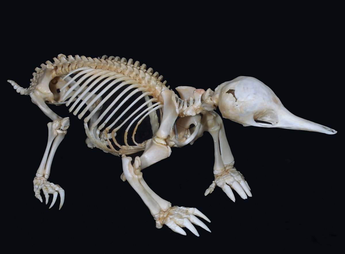
The omovertebral bone is thought to be of scapular origin, as an overgrowth of the epiphysis on the posterior border of the scapula. Amphibians have a very large suprascapular extension that is separated from the scapula by cartilage or a joint. Thornback skates have a distinct bone, the suprascapula, that is fused to the spine and articulates with the scapula. Reptiles and some early mammals (cynodonts and monotremes) have large suprascapular extensions of the scapula that project over the spine and are in close proximity to the spinous processes.

In monotremes, such as platypus (above) and echidna (below), this suprascapular extension may extend across the midline and overlie the other suprascapular extension.
Report of a patient with an omovertebral bone not associated with a Sprengel deformity has cast some doubt on the theory of a scapular origin of the bone. Other theories include a vertebral origin, as an outgrowth of the spinous process; or an origin independent of either the spine or scapula (either as an ossification of the connective tissues between the spine and scapula, or as a rudimentary scapula related to early clefting of the embryo).
References
- Azouz EM. CT demonstration of omovertebral bone. Pediatr Radiol. 2007 Apr;37(4):404.
- Williams MS. Developmental anomalies of the scapula-the "omo"st forgotten bone. Am J Med Genet A. 2003 Aug 1;120A(4):583-7.
No comments:
Post a Comment
Note: Only a member of this blog may post a comment.